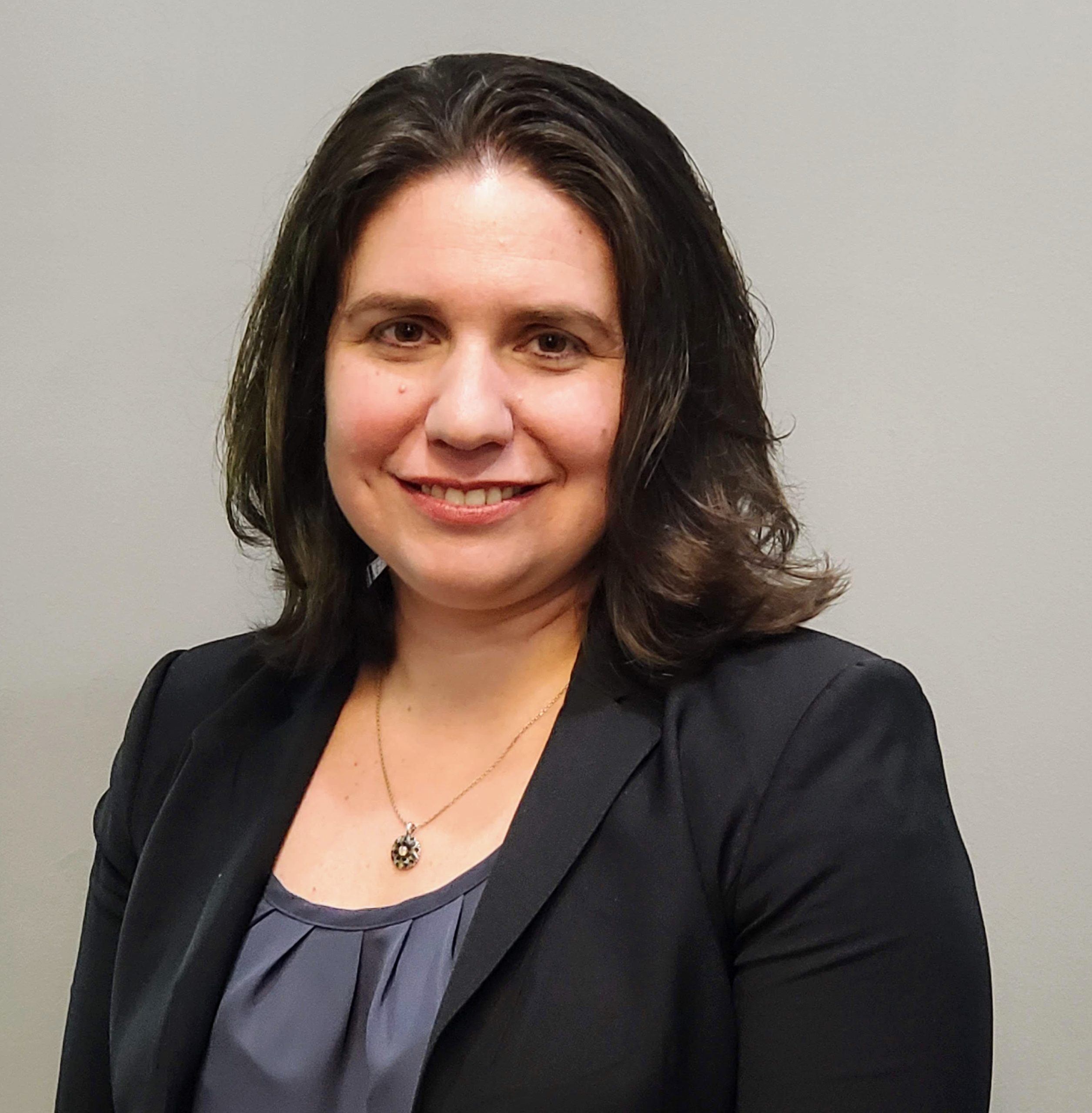
To view this, you need to install the Flash Player 5. Please go to here and download it.

twitter facebook linkedin youtube blog

Traumatic Brain Injuries New York, New Jersey, Philadelphia

Recent breakthroughs in medical technology allow our brain injury attorneys to consult local neurologists and neuropsychologists for advanced recognition of your head injuries. Until very recently, standard imaging technology could not detect injuries to the brain unless they resulted in intracranial bleeding. However, new medical technology has helped the Locks Law Firm to definitively demonstrate, in court, where injuries have occurred within the brain. These technological developments give our brain injury attorneys the ability to build stronger, more watertight cases for our clients.

If you have suffered a traumatic brain injury as the result of another party's reckless, careless, or negligent actions, you may be entitled to compensation. Proudly serving Newark, Trenton, and beyond, our Cherry Hill, New Jersey brain injury attorneys can provide more information on the following imaging devices, and inform you if using such technology would be beneficial to your claim. Contact our Pennsylvania, New York, and New Jersey personal injury lawyers for a case consultation.

Magnetic Resonance Imaging (MRI)

The MRI scanning device uses a nuclear magnetic resonance spectrometer to produce images of the inside of the brain. Utilizing the latest Tesla 3.0 magnets, the current MRI devices have a much greater chance of seeing damaged brain tissue than their predecessors. Even if there is no intracranial bleeding, the current generation of MRI machines is likely to detect brain damage if there is any. Whereas a CT scan uses ionizing radiation in the form of an X-Ray, the MRI device uses a non-ionizing radio frequency to make its images, and is best suited for non-calcified tissue, such as the soft tissue of the brain.  The Tesla 3 MRI is able to demonstrate injuries in the brain that were previously not seen on MRI films and the use of this latest technology may be critical to demonstrating the injury.

Back to top

Computed Tomography (CT) Scan

A CT scan, also known as a CAT scan, is one of the most common devices used for imaging of the brain following a head injury. A CT scan is capable of recording bleeding within the cranium, and should be used by medical facilities if there is any reason to believe that the brain may have been damaged. Though an MRI machine is able to take more precise images, a CT scan is capable of detecting hematoma, or bleeding in the brain, which could help doctors determine if there is a dangerous amount of pressure inside of the skull. Because CT scanning machines are relatively inexpensive and readily available at most hospitals, a failure by a medical staff to utilize a CT scan to diagnose a brain injury could lead to a valid medical malpractice claim.

Back to top

Positron Emission Tomography (PET) Scan

A PET scan device detects the emission of positrons in the body from a small, harmless radioactive substance administered to the patient before the procedure, usually in the form of glucose. The PET Scan is a dynamic test that measures the rate at which the brain metabolizes one of its most important fuels, glucose.  Because it measures brain activity as opposed to brain structure, it is often able to demonstrate an injury that is not seen on MRI or CT Scan.  When used in the brain, the PET Scan can identify areas of the brain that are not functioning normally. The results of a PET Scan are most useful when combined with neuropsychological test results and a medical history of the patient.

Back to top

Single Photon Emission Computed Tomography (SPECT) Scan

A SPECT scan is similar to a PET scan. It, too, involves an intravenous injection of a radionuclide into a patient. The SPECT scan then detects the photons that are emitted from the radionuclide as it circulates throughout the body. The healthy tissue in the body will absorb these radionuclides at a different rate than damaged or diseased tissue. Any indication of a deviation in the absorption of the radionuclide into the patient's tissue could point to cancerous or damaged tissue. Utilizing this technology, a doctor could detect the function and metabolism of the brain to see if it was healthy or if had been damaged by a traumatic head injury. A CAT scan and MRI scan can only look at the structure of an object, not the function.

Back to top

Diffusion Tensor Imaging

This type of imaging is an extension of magnetic resonance imaging. It works on the principle of mapping the white matter of the brain by measuring the restricted diffusion of water in tissue. The imaging device constructs tensors, or three-dimensional matrixes, of the brain that indicate the shape and direction of the water diffusion. The clinical applications of this type of imaging are in locating white-matter lesions, axonal disruption and tumors, and indicating trauma or cancer, respectively. This imaging technology gives doctors the ability to see the brain connectors, the white matter, with amazing clarity, affording them an unprecedented ability to detect brain damage.

If you are the victim of another's negligence, please contact our Philadelphia, Trenton, Cherry Hill, and Newark area brain injury attorneys and schedule a free case review.

Back to top

Functional Magnetic Resonance Imaging (fMRI)

Functional MRI is one of the most recent developments in neural imaging, and involves the detection of hemodynamic responses in the brain when subjected to stimuli. Based on the understanding that blood flow in the brain and neural activity are closely related, this new technology notes how oxygenated and deoxygenated blood flows in the brain by looking at how the blood reacts to an external magnetic field. By looking at how the brain is operating, where the oxygenated blood is flowing, and how the blood is being utilized by areas of neural activity, doctors can determine if the changes in cerebral blood flow and oxygen consumption in the brain indicate a healthy or damaged brain.

Back to top

Contact Our Brain Injury Attorneys Today

At the Locks Law Firm, our brain injury attorneys have a thorough familiarity with the advanced recognition techniques used in head injury diagnosis and are able to use the information gleaned from these tests to strengthen our clients' claims. Our experience in litigating brain injury cases has made us adept at overcoming the many hurdles associated with entering these imaging results as evidence in court.

Contact the Locks Law Firm — conveniently located to Philadelphia, eastern Pennsylvania, Trenton, Cherry Hill, and Newark, New Jersey — to schedule an appointment with our brain injury attorneys to discuss your claim. Representing victims throughout the tri-state area, all of our cases are handled on a contingency fee basis: if we don't obtain compensation in your case, we won't accept legal fees.

Back to top

If you are in the Newark or Trenton areas, contact our Cherry Hill, New Jersey office to schedule a consultation with our brain injury attorneys or to learn more about the methods used for advanced recognition of a brain injury.